Contact number: 020 7806 4030
Private Ultrasound Scans in London
Ultrasound is one of the most widely used and safest imaging techniques in medicine. It uses high-frequency sound waves to create real-time images of the inside of the body with no known side effects. At St John & St Elizabeth Hospital, we offer private ultrasound scans in London, providing fast access to high-quality diagnostics across a range of medical specialties.
Whether it’s monitoring a pregnancy, assessing abdominal pain, guiding joint injections or evaluating blood flow, diagnostic ultrasound plays a vital role in early detection and treatment planning. All scans are carried out by expert radiologists or vascular scientists using the latest ultrasound technology in a calm and supportive setting.
You’ll be looked after by our dedicated imaging team throughout your visit. Our radiologists perform and interpret most ultrasound scans, while vascular scientists are responsible for ultrasound assessments of the arteries and veins. Supporting them are our radiology department assistants, who help ensure your experience is smooth, safe and efficient.
Get peace of mind
At St John & St Elizabeth Hospital, we offer fast access to private ultrasound appointments — often available the next working day. All scans are carried out on-site by experienced specialists, with reports typically delivered to your referring clinician within 48 hours. Our patients benefit from short wait times, expert-led care, and a smooth, professional experience from start to finish. Whether you’re self-paying or insured, our team is here to guide you every step of the way.
What is the cost of an ultrasound scan?
From £435*
The price shown includes all hospital-related costs for your ultrasound It does not include consultation fees or any additional diagnostics that may be required.
*This price depends on the body part and number if images needed.
How to pay for your test
If you’re… paying for yourself
Did you know you don’t need private medical insurance to come to St John & St Elizabeth Hospital? As a self-pay patient, you can access safe, outstanding quality health care at times to suit you.
For scans and tests, as well as to see most consultants, you’ll still need to be referred by a medical professional like your GP, but as a self-pay patient, the process is more straightforward. You won’t need authorisation from an insurance provider, and you’ll have greater choice of consultant and appointment times.
If you’re… insured
St John & St Elizabeth Hospital is approved by all major medical insurance companies. If you have a personal private health insurance policy, or your company provide it for you, you can use it to pay for your care from your initial consultation through to treatment, surgery and aftercare such as physiotherapy. Not all private health insurance plans cover the same things. It’s very important to check exactly what you are covered for with your insurance provider.
Frequently asked questions about ultrasound
Once your ultrasound appointment is confirmed, you’ll receive a letter with the date, time, and location. Preparation depends on the area being scanned. You may be asked to fast or drink water to fill your bladder beforehand. If you’re taking medication, continue as normal. Wear loose clothing so the area being scanned can be easily uncovered. Our team will give you all necessary instructions ahead of your appointment.
- All patients are required to check-in and register at the imaging reception before any appointment. Please ensure you arrive early for your appointment to complete registration.
- Self-Funding Patients: The imaging administrative team can provide quotes for all examinations in the imaging department, if you would like to know the cost of your examination before your appointment, please contact us.
- Insured Patients: If you are insured please bring your insurance membership number and authorisation number with you to your imaging appointment. If you do not provide these details at the time of registration you may be asked to self-fund for the exam.
- A copy of your report will be sent to your referring doctor within 48 hours of your appointment. If you are seeing your doctor within 48 hours please let one of the imaging team know so we can expedite this.
- We are unable to release imaging reports to you, as the patient until your doctor has had the opportunity to discuss the results with you. You doctor should be able to provide you with a copy of the report, please contact us following your consultation if they are unable to do so
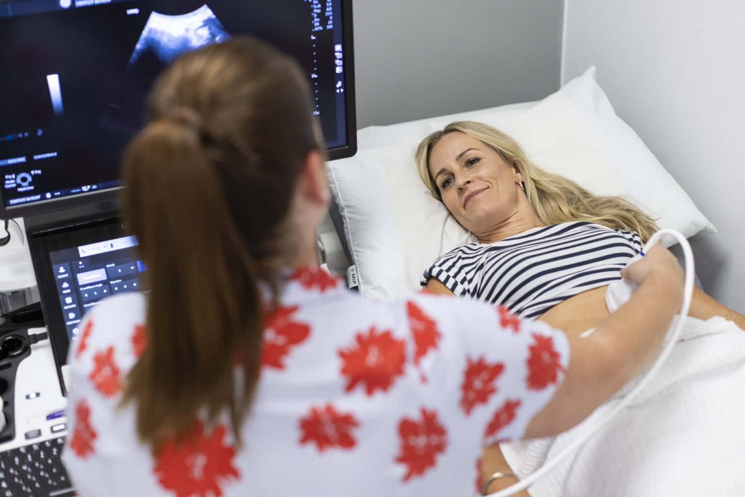
When you arrive, you’ll be guided to the scan room. You’ll be asked to remove clothing from the area being examined and lie on the examination couch. A clear gel will be applied, and the radiologist will move a handheld device (transducer) over the skin to capture images. The room lights may be dimmed to help the radiologist see the screen clearly. Once the scan is complete, you’ll be given tissues to wipe off the gel and can get dressed straight away.
The length of the scan depends on the part of the body being examined and the detail your referrer has requested. Most scans take between 10 and 30 minutes.
Ultrasound itself is painless, but you may feel slight pressure when the radiologist presses the probe to get clearer images. If you feel any discomfort, let the radiologist know straight away.
You can leave the department immediately after your scan and return to your normal activities, including eating and drinking. Your images will be reviewed by a radiologist, and a report will be sent to your referring doctor, who will then discuss the results with you and advise on any next steps.